Why Lung Health Matters on World MS Day – And Every Day
Each year on May 30, the world unites to recognise World MS Day – a powerful movement that brings together the global Multiple Sclerosis (MS)
Previously we had assumed that PEP and OPEP devices (like AirPhysio) would help improve the effectiveness and distribution of medication due to the fact that they clear mucus and lung expansion, allowing improved distribution of the aerosol medication and better access to the inflamed airways in the lungs.
There are, however, five studies available which show the benefits of improved distribution and effectiveness of aerosol medication when combined with the use of PEP, OPEP and Pressure Support devices.
The five medical studies include four different medical studies with the use of bronchodilators and PEP devices, and 1 study with Pressure Support through different aerosol distribution methods:
This study assessed the effectiveness of a positive expiratory pressure (PEP) device on beta2-agonist (short term reliever) nebulization therapy by measuring the pulmonary function before and after nebulization therapy in 54 asthmatic patients.
The results show that the use of PEP device after beta2-agonist nebulization therapy improved pulmonary function compared with the use of beta2-agonist nebulization therapy alone, as shown by the increases in forced mid-expiratory flow and forced vital capacity (FVC).
Patients with forced expiratory volume in 1 sec (FEV1) below 85% FVC obtained a significant improvement in FEV1 and FVC after using the PEP device.
When the PEP device was used before beta2-agonist nebulization therapy, there were no obvious direct bronchodilatory effects, but the use of PEP device after beta2-agonist therapy showed significantly enhanced the bronchodilatory effect of beta2-agonist therapy in patients with an FEV1 below 85% FVC.
Conclusion
The use of PEP device after the use of beta2-agonist shows a significantly enhanced effect of bronchodilative effect in patients with an FEV1 below 85% FVC, over beta2-agonist therapy by itself.
It was put down as this outcome of the additional effect of the PEP device use in improving pulmonary function after beta2-agonist nebulization therapy might be a result of an enhancement in mucus clearance.
Abstract – Effectiveness of a positive expiratory pressure device in conjunction with beta2-agonist nebulization therapy for bronchial asthma. – PubMed – NCBI
Full Article – Effectiveness of a positive expiratory pressure device in conjunction with β2-agonist nebulization therapy for bronchial asthma
This randomized, double-blinded study was carried out to differentiate the effect of heliox (helium and oxygen) and oxygen with and without positive expiratory pressure (PEP), on delivery of radiotagged inhaled bronchodilators on pulmonary function and deposition in asthmatics.
They chose 32 patients between the ages of 18 and 65 years who were diagnosed with stable moderate to severe asthma, and they were randomly assigned into four groups: (1) Heliox + PEP (n = 6), (2) Oxygen + PEP (n = 6), (3) Heliox (n = 11) and (4) Oxygen without PEP (n = 9).
Both gas type and PEP level were blinded to the investigators. Images were acquired with a single-head scintillation camera with the longitudinal and transverse division of the right lung as regions of interest (ROIs).
While all groups responded to bronchodilators, only group 1 showed an increase in Forced Expiratory Volume in 1 second (FEV1) %predicted and Inspiratory Capacity (IC) (the amount of air that can be inhaled after the end of a normal expiration) compared to the other groups (p < 0.04).
When evaluating the ROI in the vertical gradient, we observed higher deposition in the middle and lower third in groups 1 (Heliox + PEP) (p = 0.02) and 2 (Oxygen + PEP) (p = 0.01) compared to group 3 (Heliox).
In the horizontal gradient, a higher deposition in the central region in groups 1 (Heliox + PEP) (p = 0.03) and 2 (Oxygen + PEP) (p = 0.02) compared to group 3 (Heliox) and intermediate region of group 2 (Oxygen + PEP) compared to group 3 (Heliox).
The increase in IC could be explained by the physical characteristics of heliox, which allows the formation of less turbulent airflow, thereby generating more flow and time during expiration, leading to a reduction in dynamic hyperinflation and increase in IC. On the other hand, the use of PEP prevents airway collapse during expiration, decreases expiratory resistance, prolongs expiratory time and reduces the intrinsic positive end-expiratory pressure (PEEPi),8 promoting an increase in IC.
A significant increase in FEV1 was observed in the heliox þ PEP group, which did not occur in other groups. Our findings can be correlated with those of Tsai et al. .28 (document mentioned above) who assessed 54 stable asthmatic patients before and after nebulization with bronchodilators associated with PEPP and observed improvement with respect to FEV1, PEF, FVC, as well as improvement in mucociliary clearance.
Conclusion
They concluded that aerosol deposition was higher in groups with PEP independent of gas used (i.e. both oxygen and heliox), while bronchodilator response with heliox + PEP improved FEV1 % and IC compared to administration with Oxygen, Oxygen + PEP and Heliox alone.
Abstract – https://www.ncbi.nlm.nih.gov/pubmed/23664767
Evaluation of lung function and deposition of aerosolized bronchodilators carried by heliox associated with positive expiratory pressure in stable … – PubMed – NCBI
Full Article – Evaluation of lung function and deposition of aerosolized bronchodilators carried by heliox associated with positive expiratory pressure in stable … – PubMed – NCBI Evaluation of lung function and deposition of aerosolized bronchodilators carried by heliox associated with positive expiratory pressure in stable asthmatics_ A randomized clinical trial
Study objective: To determine whether the use of a mucus clearance device (Flutter OPEP device) could improve the bronchodilator response to inhaled ipratropium and salbutamol delivered by a metered-dose inhaler in patients with stable, severe COPD.
Patients: Twenty-three patients with severe COPD were studied. Mean +or- SD age was 71.7 +or- 6.3 years. Mean FEV1 was 0.74 +or- 0.28 L or 34.5 +or- 12.7% predicted.
Methods: Patients were tested in random order on two subsequent days after using a mucus clearance device or a sham mucus clearance device (ball bearing removed from the device). A bronchodilator (four puffs; each puff delivering 20 ug of ipratropium bromide and 120 ug of salbutamol sulphate) was administered by metered-dose inhaler with a holding chamber after use of the mucus clearance device or sham mucus clearance device. Spirometry was performed before and after use of the mucus clearance device or sham mucus clearance device, and at 30 min, 60 min, and 120 min after the bronchodilator. Six-minute walk distance was tested between 30 min and 60 min; oxygen saturation, pulse, and a dyspnea score were recorded before and after walking.
Results: Immediately after use of the mucus clearance device, but not the sham mucus clearance device, there was a statistically significant (p < 0.05) improvement in FEV1 and FVC (11 +or- 24% vs 1 +or- 7% and 18 +or- 33% vs 6 +or- 18%, respectively). Whether patients were pretreated with the mucus clearance device or sham mucus clearance device, there was a significant improvement in FEV1 and FVC compared to baseline with combined bronchodilator therapy. At 120 min, the change in FEV1 after treatment with the mucus clearance device was greater than with the sham mucus clearance device (186 +or- 110 mL vs 130 +or- 120 mL; p < 0.05). When comparing the mucus clearance device to the sham mucus clearance device, 6-min walk distance was greater (174 +or- 92 m vs 162 +or- 86 m; p < 0.05), with less dyspnea before and at the end of walking.
Conclusion: Patients with severe COPD may demonstrate a significant bronchodilator response to combined ipratropium and salbutamol delivered by a metered-dose inhaler. This response may be enhanced and additional functional improvement obtained with the prior use of a bronchial mucus clearance device.
Use of a Mucus Clearance Device Enhances the Bronchodilator Response in Patients with Stable COPD – selected study
The influence of positive expiratory pressure (PEP) applied during inhalation of a beta 2-agonist in treatment of bronchial asthma was investigated in a randomized crossover study with two-week treatment periods.
In one period, two puffs (0.5 mg) of terbutaline was given from a metered-dose inhaler and inhaled through a device consisting of a cone spacer connected to a facemask giving PEP (10 to 15 cm H2O).
In a second period, terbutaline 0.5 mg was inhaled similarly but without PEP, and in a third-period placebo spray was inhaled with PEP.
Treatments were given three times daily. Peak expiratory flow (PEF) was measured before and after each inhalation and symptom scores for dyspnea, cough, and mucus production were noted in a diary.
All treatments increased PEF significantly (p less than 0.0001). The mean increase was 32 L/min during treatment with terbutaline and PEP.
This was greater than the increase of 25 L/min during terbutaline treatment (p = 0.005).
The increase in PEF during terbutaline treatment was significantly higher than the achieved 18 L/min during PEP (p = 0.026).
Conclusion
The study showed improved bronchodilation when PEP was combined with inhalation of beta 2-agonist compared with beta 2-agonist alone.
Abstract – https://www.ncbi.nlm.nih.gov/pubmed/186410
Full Article – https://www.sciencedirect.com/science/article/abs/pii/S0012369216371550
INTRODUCTION:
Positive expiratory airway pressure seems to dilate narrowed or collapsed airways, but this may be accompanied by a maintained and harmful increase in resting lung volume in obstructive pulmonary disease.
PURPOSE:
To evaluate the influence of inhaled terbutaline and positive expiratory pressure (PEP) on airway resistance (Raw) and functional residual capacity (FRC) in bronchial asthma.
DESIGN:
Randomized crossover design, single-blind with regard to inhaled medication, open with regard to PEP (PEP can be felt).
MATERIAL AND METHODS:
Ten patients with bronchial asthma inhaled placebo and terbutaline in doses of 0.125 mg, 0.5 mg, and 1.5 mg by cone spacer combined with a facemask giving 0, 10, or 15 cm H2O PEP on separate days. FRC and Raw were measured by body plethysmography before and after inhalations. Data were analyzed by analysis of variance with terbutaline dose and PEP as factor levels.
RESULTS:
The effect of terbutaline: Raw decreased significantly (p < 0.0001) after 0.125 mg and 1.5 mg. The FRC did not change significantly. The effect of PEP: Raw decreased, but significantly only when the dose of 1.5 mg terbutaline was excluded from the analysis. Raw decreased with PEP 10 and 15 cm H2O, mean 0.6 (95 percent CI: -1.1, -0.2) and 0.9 (95 percent CI: -1.3, -0.4) cm H2O/L/s. The FRC did not change significantly with the PEP level.
CONCLUSION:
PEP only had an influence on Raw when insufficient doses of terbutaline were inhaled, whereas once an efficient dose of terbutaline was administered, significant bronchodilation was achieved with or without PEP. Positive expiratory pressure did not increase FRC.
Abstract – https://www.ncbi.nlm.nih.gov/pubmed/8404176
Nebulized aerosols are commonly used to deliver drugs into the lungs of patients with cystic fibrosis (CF).
The aim of this study was to assess the effectiveness of pressure-support (PS) ventilation in increasing aerosol deposition within the lungs of children with CF.
An in vitro study demonstrated the feasibility of coupling a breath-actuated nebulizer to a PS device. An in vivo study was done with 18 children (ages 6 to 21 yr) with clinically stable CF, each of whom underwent both a standard and a PS-driven ventilation scan (control session and PS session, respectively).
In addition, a perfusion scan was used to determine lung outlines and to construct a geometric model for quantifying aerosol deposition by radioactivity counting in MBq.
Homogeneity of nebulization was evaluated from the four first-order moments of aerosol distribution in the peripheral and central lung regions. The time-activity nebulization curve was linear in all patients, with higher slopes during the PS than during the control session (0.43 +/- 0.07 [mean +/- SD] MBq/min and 0.32 +/- 0.23 MBq/min, respectively; p < 0.018).
Quantitatively, aerosol deposition was about 30% greater after the PS session (4.4 +/- 2.7 MBq) than after the control session (3.4 +/- 2.1 MBq; p < 0.05).
Similarly, deposition efficacy (as a percentage of nebulizer output) was significantly better during the PS session than during the control session (15.3 +/- 8.3% versus 11.5 +/- 5.7%, p < 0.05).
No differences in the regional deposition pattern or inhomogeneity of uptake were observed.
Conclusion
In conclusion, our data show that driving the delivery of a nebulized aerosol by noninvasive PS ventilation enhances total lung aerosol deposition without increasing particle impaction in the proximal airways.
Abstract – https://www.ncbi.nlm.nih.gov/pubmed/11112150
Full Article – https://www.atsjournals.org/doi/full/10.1164/ajrccm.162.6.2003069
Based on these studies, the evidence indicates that by combining the use of a PEP or OPEP device with aerosol medication and therapies, the effect and distribution of the aerosol and gases are improved. They also state that bronchodilative effect and response appears to be improved through the combined use of PEP and OPEP device with gas and aerosol treatments and therapies.
Each year on May 30, the world unites to recognise World MS Day – a powerful movement that brings together the global Multiple Sclerosis (MS)
Asthma is more common than many people realise — affecting over 262 million individuals around the world. Yet despite how many lives it touches, asthma
In the pursuit of optimal respiratory health, understanding and implementing techniques like Chest Percussion can be a game-changer. This method, designed to enhance lung function
AirPhysio may be able to help by assisting with
✅Clearing the mucus clogging airways
✅Helps Promote Optimal Lung Capacity
✅Helps Promote Optimal Lung Hygiene
✅Helps treat Respiratory Conditions
✅May Reduce the chance of developing lung infections
✅Recommended by doctors, physical Therapists & Pulmonologists
✅Helps Make Breathing Easier
✅Sold in 106 countries globally


Do you suffer from Dry or Persistent Cough, Breathlessness, Respiratory or Severe Respiratory Conditions, or Shortness of Breath as you Get Older?

THIS PRODUCT MAY NOT BE RIGHT FOR YOU. READ THE WARNINGS BEFORE PURCHASE (Contraindications of Untreated pneumothorax; Tuberculosis; Oesophageal surgery; Right-sided heart failure; Middle ear pathology, such as ruptured tympanic membrane)
FOLLOW THE DIRECTIONS FOR USE
IF SYMPTOMS PERSIST, TALK TO YOUR HEALTH PROFESSIONAL

Gallery
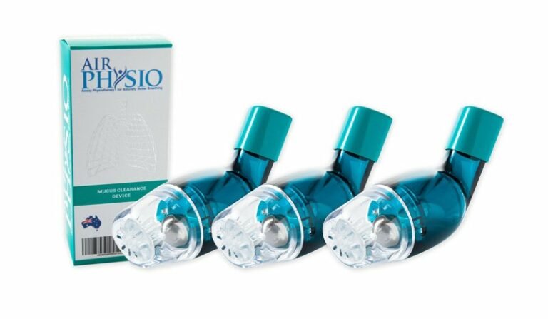
AirPhysio is an Oscillating Positive Expiratory Pressure (OPEP) device that is used for mucus clearance and lung expansion to help in the treatment of respiratory conditions. The use of the AirPhysio device helps to facilitate secretion mobilization and maximize lung volume for cleaner healthier lungs.
© AirPhysio 2022 – All Rights Reserved
| Cookie | Duration | Description |
|---|---|---|
| cookielawinfo-checkbox-analytics | 11 months | This cookie is set by GDPR Cookie Consent plugin. The cookie is used to store the user consent for the cookies in the category "Analytics". |
| cookielawinfo-checkbox-functional | 11 months | The cookie is set by GDPR cookie consent to record the user consent for the cookies in the category "Functional". |
| cookielawinfo-checkbox-necessary | 11 months | This cookie is set by GDPR Cookie Consent plugin. The cookies is used to store the user consent for the cookies in the category "Necessary". |
| cookielawinfo-checkbox-others | 11 months | This cookie is set by GDPR Cookie Consent plugin. The cookie is used to store the user consent for the cookies in the category "Other. |
| cookielawinfo-checkbox-performance | 11 months | This cookie is set by GDPR Cookie Consent plugin. The cookie is used to store the user consent for the cookies in the category "Performance". |
| viewed_cookie_policy | 11 months | The cookie is set by the GDPR Cookie Consent plugin and is used to store whether or not user has consented to the use of cookies. It does not store any personal data. |